Illuminating Cell Division with AI

Step into the lab of Orlando Arguello-Miranda, assistant professor in the Department of Plant and Microbial Biology at NC State University, and you’ll find a bustling hub at the intersection of microbiology and computer science focused on using algorithms to understand the molecular networks that drive cell division.
The lab brings biologists and computer scientists together to gain new insight into biological processes. It’s an interdisciplinary approach to innovation that few laboratories explore in depth; however, Miranda has always focused on combining different scientific disciplines to answer biological mysteries.
After receiving his Ph.D. in biochemistry from the Technische Universität Dresden in Germany, Miranda began working with image processing algorithms as a postdoctoral researcher in 2016 at UT Southwestern Medical Center. At the time, artificial intelligence (AI) applied to cell biology was still viewed as a trend that would fade with time. But Miranda was particularly interested in how new AI algorithms could understand cell division. Receiving the K99 Pathway to Independence Award from the National Institutes of Health gave him the opportunity to focus on that research full-time.
Since joining the Department of Plant and Microbial Biology as part of the Quantitative and Computational Developmental Biology Cluster, Miranda has taken his research to the next level, developing new sensors to track intracellular processes and engineering experimental setups for observing entire signaling pathways in cells.
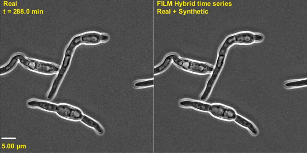
“We have accelerated the analysis of living cells to a point we only could dream of four years ago,” he says. “By combining generative AI and live cell microscopy, we are now able to track individual cells and microorganisms without interruption throughout their entire lives, including cell division, reproduction and dormancy. By contrast, previous approaches could only study one process at a time.”
Viewing Microbial Life Cycles

One ongoing project in Miranda’s lab has led to the development of an algorithm called FIEST (Frame Interpolation Enhanced Single-cell Tracking) that tracks individual cells and their growth and division throughout their entire life cycle, including their descendants. This approach, powered by a combination of AI tools, can follow individual cells even when they completely change size or shape, for instance, when exposed to harsh conditions (such as a low nutrient environment) and during sexual reproduction, which has always been a challenge for single-cell cell biology.
Originally developed in yeast, the lab has continued to refine the algorithmic AI procedure to track and count other cells, including bacteria, cancer cells and human organoids. An earlier version of the algorithm was published as an Open-source Python package that has been downloaded over 35,000 times.
“The first time we saw the FIEST algorithm in action, we all shouted out of excitement. We had a very complicated video where hundreds of cells reproduced in a chaotic manner, and all other tracking algorithms had failed at this point,” Miranda recalls. “We developed the FIEST algorithm out of need, but did not know whether it would work.
“However, when the results came back, there was a moment of silence, followed by shouting. Every single one of the 632 cells had been tracked correctly, regardless of their shape, size or movement.”
The most recent publication from the Miranda lab showcases how the lab’s algorithms have grown and improved over time. The algorithms were able to image and track yeast cells through three generations of growth and reproduction, a feat once thought impossible because it is equivalent to recording the lives of grandparents, parents and children, three complete generations, in video.
As word spread about the algorithms’ capabilities, Miranda recalls countless emails and conversations from other labs requesting help with image analysis. He quickly realized this was not work for a research lab, but a service-based company.
To address this need, Miranda has co-founded ilustris, a platform for image analysis, with Kevin Flores, an NC State mathematics professor. The company’s name derives from ancient Latin, meaning “bright” or “lustrous,” representing their desire to illuminate data in a new light. Labs can easily produce thousands of microscopy images, but face a bottleneck when analyzing them. Through its platform, Ilustris will allow users to upload their images and use the lab’s algorithms to rapidly track and analyze microscopy images.
Strawberries in the Spotlight
The Miranda Lab is already developing another AI-powered imaging tool to support strawberry farmers in North Carolina.
In 2018, a new pathogen, Neopestalotiopsis, began infecting strawberry plants across the Southeast. By 2022, it had spread to North Carolina strawberry farms, causing crown, leaf and fruit rot, ultimately killing many plants. There are no resistant varieties available on the market, and the best way to manage the disease is to avoid bringing infected nursery plants to the farm.
Fast facts about the strawberry industry in North Carolina:
- North Carolina ranks third nationally in strawberry production.
- NC State has a strawberry breeding program led by Gina Fernandez in the Department of Horticultural Science, and plant pathology and disease management programs led by Tika Adhikari and Frank Louws, in the Department of Entomology and Plant Pathology.
Miranda is working together with farmers in New Hanover County and Tika Adhikari, a principal research scholar in the Department of Entomology and Plant Pathology, to explore the use of AI-powered microscopy techniques to rapidly identify fungal contamination, such as Neopestalotiopsis spores, in fields and nurseries. The spores of this pathogen have a unique shape, “like bananas with party hats,” says Miranda, making them easy for AI to identify in an image.
The speed of image processing is a huge advantage in comparison to traditional techniques, which require complex identification kits and often can not be deployed in the field. Image-based detection methods will allow farmers to respond to pathogens without waiting for lab results, and instead visually and directly confirm the presence of a pathogen.
An additional benefit of this method is the ability to determine whether the pathogen is alive. While a standard chemical test detects the presence of a pathogen’s DNA, it cannot tell farmers if the pathogen is alive or if it is a trace of DNA left behind by a destroyed cell or other form of contamination. Image analysis, however, looks at the cells’ features, giving the computer program a better understanding of whether the pathogen is alive or not and what stage of the life cycle it is in.
Future-Focused
While launching Ilustris, Miranda continues to grow the use of AI to understand biochemical processes within cells. He’s expanding his work further, with current projects focused on the use of generative AI to distinguish proteins inside living cells.
Miranda sees AI as the tool that will allow humankind to tackle the complexity of biological systems.
“There are millions of multiple proteins at any given time in a single cell interacting and directing the process of cell division,” he says. “Using AI, I envision we will eventually understand each molecule and each interaction in living cells, which will give us a better understanding of diseases and how to improve agriculture.”
Miranda credits Adhikari and Flores for their crucial collaborations with his lab. A National Science Foundation grant from the Emerging Mathematics in Biology program in 2024 supported Miranda and Flores’ work. They continue to collaborate on their project, which relies on accurate high-throughput tracking and quantification of protein expression in live cells.
With advanced imaging techniques, Miranda envisions a future in which researchers can track every protein and structure within a living cell, revolutionizing how scientists produce information to improve human life.


